
3D Cell Explorer-fluo
Combine high-quality tomographic data with fluorescent markers.
Combine high-quality tomographic data with fluorescent markers.
Why the 3D Cell Explorer-ƒluo?
Combine high-quality RI tomographic data and fluorescence markers
You want to extend your understanding of cell structures and cell mechanisms? Thanks to our 3D tomographic technology you can now have a powerful platform that combines RI information with fluorescence markers.
Go beyond fluorescence limitation
You are frustrated by the limitations of fluorescence markers? You would like to go further? Combine them with Nanolive’s digital staining and push the limits forward.
Achieve specific cell analysis while preserving cell life
Forget about fixation! With the 3D Cell Explorer-fluo you can now observe your cells as they are: alive, 3D and in motion!
Perform infinite live cell imaging
Limit cell damages caused by fluorescence markers, bleaching, and phototoxicity. The 3D Cell Explorer-fluo allows you to transform your chemical markers into physical ones which can be monitored for a limitless amount of time.
COMPLETE 3D SOLUTION
Combine high-quality tomographic data with fluorescent markers
MULTIPLEXING
Explore up to 10 markers in parallel
EXTENDED LIVE CELL IMAGING
Limit cell damages caused by fluorescent markers, bleaching and phototoxicity
| Technical Features | 3D Cell Explorer-fluo |
| Illumination Source | Holotomography: Class 1 laser low power (λ=520 nm, sample exposure 0.2 mW/mm²) Fluorescence: High speed switchable <100 µs, Lifetime >20'000 hours each channel |
| Resolution | Holotomography: x,y: 200 nm; z: 400 nm (3D image) Fluorescence: rx,y: ~ 400 nm (2D image) |
| Field-of-view | Holotomography: 90 x 90 × 30 μm Fluorescence: 90 × 90 μm |
| Microscope Objective | Dry objective / 60x magnification / NA 0.82) |
| Channels | Holotomography: Up to 7 simultaneous Fluorescence: DAPI + FitC + TritC | FitC + TritC + Cy5 | DAPI + FitC + TritC / Cy5 |
| Imaging | Holotomography: 3D Fluorescence: 2D 4D time lapse: (RI + fluo) |
| Time resolution | Holotomography: 0.5 fps 3D RI frame Fluorescence: 3 fps each channel |
| Camera | USB 3.0 CMOS Sony IMX174 sensor / Quantum Efficiency (typical) 70 % (at 545 nm)/ Dark Noise (typical) 6,6 e¯ / Dynamic Range (typical) 73,7 dB |
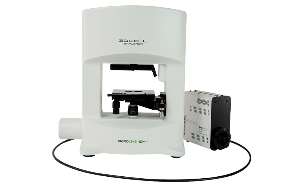
| Dimensions (width x depth x height in mm) |
3D Cell Explorer-fluo: 380 x 170 x 445 Fluorescence module: 77 x 186 × 162 |
| Weight | 12 kg |